Under the Microscope
Richard Bell’s Wild West Yorkshire nature diary, Sunday, 20th September 2009
previous | home page | this month| e-
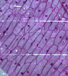
To get you started there are 5 prepared slides, plus 7 blanks. I was amazed how clearly I could see the cell structures in a prepared section of onion stem (right).
Also included is an experiment for rearing brine shrimps from eggs supplied with the kit, using a little sea salt, also supplied. The instructions warn that ‘The brine shrimp are not suitable for eating!’
You can illuminate from above the subject or below or in combination and I found
that a desk-
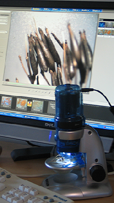
Traveler USB Microscope
Aldi, August offer price, £29.95
with 10x, 60x, 200x magnification
1.3 megapixel camera
For
making photos and recording video clips
2 intergrated light sources
Ulead VideoStudio
7 software
Website: Traveler
x 10
x 60
x 200
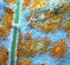
WHILE CUTTING back vegetation around the pond I came across some coltsfoot leaves
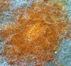
spotted with orange coltsfoot rust fungus Coleosporum tussilaginis, which I drew
in my Rough Patch garden sketchbook. I brought it in to take a closer look using
my new Traveler USB-
On screen the feltlike hairs on the back of the leaves look like our feathery cavity wall insulation. You can imagine my surprise while looking at this alien landscape when a red plant mite trundled along the stem on the left. With a click of the mouse I was able to capture it on video.
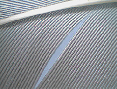
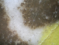
Gull wing feather, which I picked up at Langsett Reservoir last week.
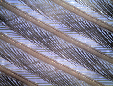
Another feather from Langsett, probably a grouse breast feather, showing the barbules, which zip together when the bird preens itself.
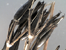
Umbellifer seeds (please let me know if you can identify which species) from the edge of a track in Thetford Forest. Taken in natural light.
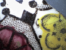
Fragment of pottery picked up in a field, probably Victorian, 10x.
Close up of the fragment at 200x. The scratches and cracks on the surface of the glaze and the bubbles in it remind me of ice.
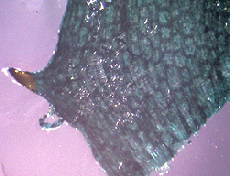
One of the prepared slides of Esthwaite Waterweed, Hydrilla verticillata, which is very rare as a native in Britain but naturalised and invasive in the USA.
Leaves minutely toothed on their margins
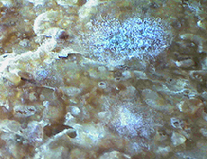
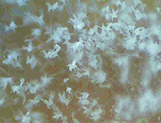
This looks like the surface of one of the moons of Jupiter but in fact it’s bread mould on an old crust seen at 10x.
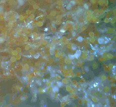
At 200x the bread mould looks like little white daffodils, each on a slender stalk. The rust mould at the top of the page looks like pins.
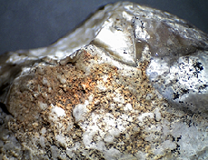
Finally a close up of a stone from my friends Rheba and Farris in Texas, that, according to a palaeontologist, was a gastrolith; a stone swallowed by a dinosaur to aid digestion.
I think this microscope deserves a place on my desk because it’s so versatile. When I had my first (and come to think of it only) full time job, working as a background artist on the film Watership Down in London in the 1970s, I used most of one week’s wages to buy a serious compound microscope from one of the technical stores on the Tottenham Court Road but I’ve never used it regularly. Having this plugged in to my computer means that it’s there for everyday use.
The focusing is simple but effective; you use the focus dials to move the stage up
or down. It’s not of course as smooth as my heavy metal compound microscope but,
as you can see, it works fine. Most fossils and rocks won’t fit on the stage under
the microscope so my solution -
previous | home page | this month| e-